Management of the Posterior Maxilla in the Compromised Patient:
Historical, Current, and Future Perspectives
CONTINUED (Page 11)
FIGURES (Page 1 of 2)
Thomas J. Balshi & Glenn J. Wolfinger
Periodontology 2000, Vol 33, 2003, 67-81.
Figure 1
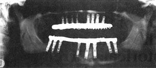
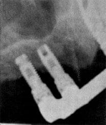
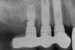
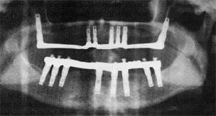
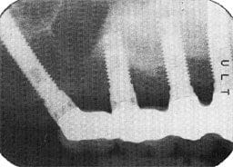
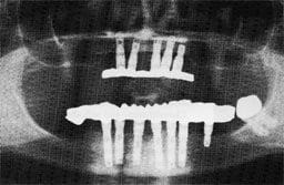
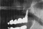
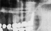
(a) Panoramic and periapical radiographs of maxillary fixed detachable prosthesis with cantilever illustrating advanced bone loss on posterior fixture.
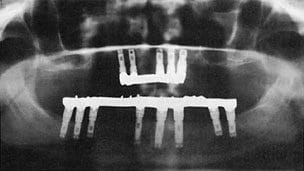
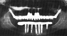
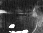
(b) Transition from fixed detachable prosthesis to maxillary implant overdenture one five implants after removal of three posterior implants with advanced bone loss. Note the bone loss on the remaining implants as well.
(a1)
(a2)
(b1)
(b2)
(b)
Figure 2a
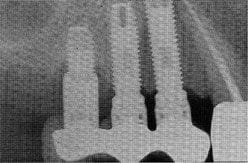
Unilateral maxillary posterior implant reconstruction showing advanced bone loss around distal fixture and subsequent implant fracture.
Figure 1c
Additional fixtures placed in the pterygoid region for extension of the overdenture bar for better stability.
Figure 2b
Replacement of fixed prosthesis without cantilever after distal implant was resurfaced.
Figure 2c
Additional implants placed for better stability of unilateral maxillary posterior prosthesis.
Figure 3a
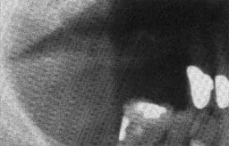
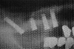
Preoperative panoramic view of advanced bone loss in maxillary posterior region.
Figure 3b
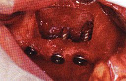
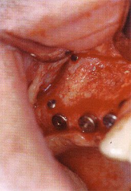
Caldwel-luc procedure in fracturing buccal plate of bone and placement of three fixtures to support and elevate the bone plate and sinus membrane.
Figure 3c
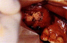
Placement of 50/50 mixture of autogenous bone and Bio-Oss bovine bone material for sinus grafting.
Figure 3d
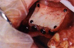
Placement of a Bio-Gide resorbable membrane with the use of four titanium tacks.
Figure 3e
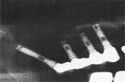
Postoperative panoramic radiograph showing the placement of three implants in the grafted area and a pterygoid fixture for posterior support.
Figure 3f
Clinical view five months postop showing complete bone fill.
Figure 3g
Panoramic view of final prosthesis in place.
(h1)
Figure 3h
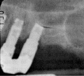
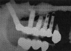
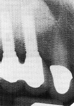
Periapical views showing final prosthesis in place and bone graft one year after surgery.
(h2)
Figure 4b
After loss of one of the anterior implants, implants were placed in the pterygoid maxillary region to support a full arch maxillary fixed detachable porcelain fused to gold prosthesis.
Figure 4a
Five implants to support a mandibular implant overdenture in the anterior region.
Figure 5c
Panoramic view thirteen years later showing the response of the restoration of an implant in the pterygomaxillary region connected to a natural tooth.
