Management of the Posterior Maxilla in the Compromised Patient:
Historical, Current, and Future Perspectives
CONTINUED (Page 12)
FIGURES (Page 2 of 2)
Thomas J. Balshi & Glenn J. Wolfinger
Periodontology 2000, Vol 33, 2003, 67-81.
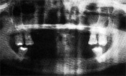
Figure 6a
Preoperative panoramic view of patient with congenitally missing teeth.
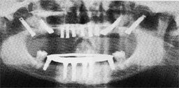
Figure 6b
Maxillary anterior implants. A total of 10 implants: 2 zygomatic and 2 in the pterygomaxillary area.
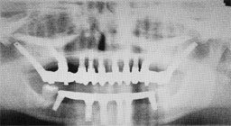
Figure 6c
Restoration of the maxillary arch utilizing implants in both the zygoma and pterygoid regions.
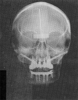
Figure 6d
Anterior/posterior cephalometric radiograph showing projection of implants in the zygoma area.
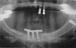
Figure 7a
Preoperative panoramic radiograph with old implant restoration.
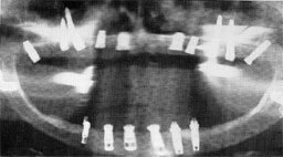
Figure 7b
Post-surgical radiograph showing implants in the severely resorbed maxilla; a total of two implants on each side were placed in the zygoma region.
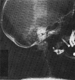
Figure 7c
Lateral cephalometric radiograph illustrating the implants in the pterygomaxillary region and the four implants in the zygoma region.
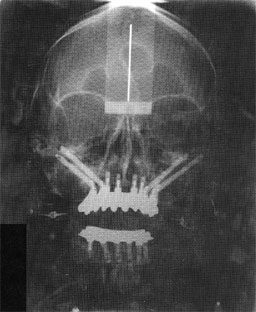
Figure 7e
Anterior/posterior cephalometric radiograph showing projection of the four implants in the zygoma region to support the full fixed maxillary reconstruction.
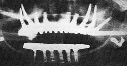
Figure 7d
Post-treatment panoramic radiograph showing reconstruction of severely resorbed maxillary utilizing the four implants in the zygoma region.